Lisa Dick
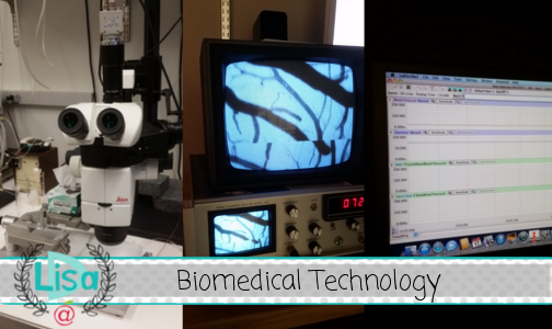
Wednesday marked a lot…A LOT of firsts for me, and yes it involved each component of this post: bodies, rats, and technology. Before you decide this is just too over the top for you…just bare with me!
Saying Dr. Michelle Turpin, one of the instructors in our Science/Math Workshop, is passionate about science would be an understatement! She LOVES science:) Wednesday marked a field trip to the Biomedical Research building at LSU Health in Shreveport. Though I didn’t know really what to expect (other than cadavers), I was happy to spend the morning out and about instead of in a classroom (I know just as bad as kids LOL).
After an overview, my group went to tour various labs. Ok, so these labs are not what you see on tv, but it was pretty neat. Dr. Turpin was about to take us to look at cells and such when she received word that we could see something even cooler. This was when the tour turned from neat to totally awesome!
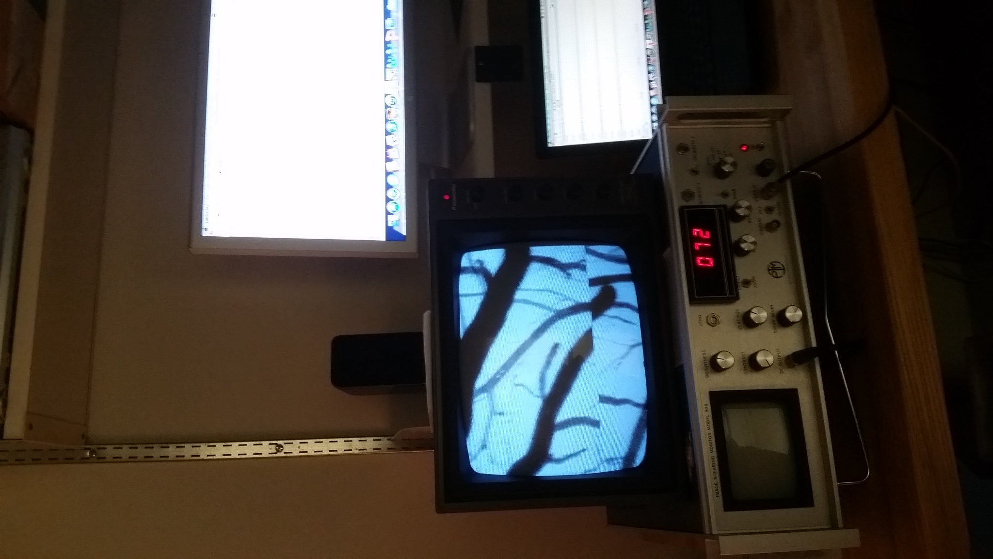 Though I am not a science guru, I totally love technology and am a data nerd! This image shearing monitor is hooked up to various data monitors and a microscope. This doctor is researching diabetes and other factors effects on strokes. The veins you are viewing in this monitor is a live rat brain! I might need a disclaimer here LOL I am referring to it as a rat, but they call it a mouse. When I think of a mouse, I draw a picture in my mind of a cute tiny creature twitching it’s nose:) This was much larger than my mental picture….but I’ll start referring to it as they did, mouse (big mouse).
The mouse was asleep and had a window in it’s head. The microscope in the main blog image is the exact microscope it was under (against policy to take picture of it). Anything they wish to observe is easily not only viewed but also an abundance of data collected in a spreadsheet/database type program.
You might notice that the monitor appears to have a “glitch” with a vertical line running through it. That is actually a key aspect of the image shearing monitor, the ability to view a previous frame to compare to the current one.
I honestly wasn’t sure if I would be able to handle the next stop, cadaver lab. Though it was a day off for the current students, it appeared as if they were all in class. The room was filled with groups of students working on their cadaver. This wasn’t anything like what I thought it would be and was actually quite interesting, but more importantly an education that just can’t be matched in another form, though our next stop came close!
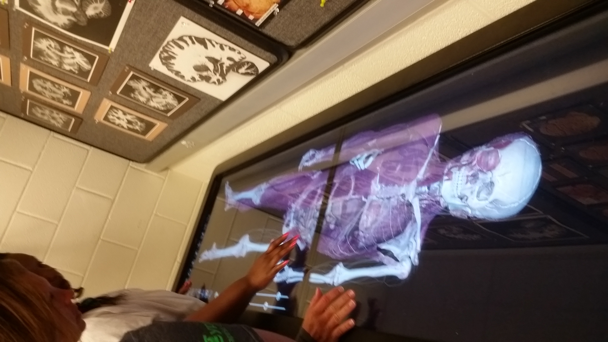
The Anatomage Table reminds me of the Smart Table on steroids Promethean ActivPanel on Full Tilt Stand. This table is as close as you can get to an actual cadaver lab and had many other features that wouldn’t be possible in the other setting such as xrays, importing various scans to the table for 3d viewing, unique dissections, and more. Here is an awesome Standford video that gives you a great overview https://www.youtube.com/watch?v=6FFd6VWIPrE
I can’t wait to add various new information and equipment to my units! What ways have you discovered to integrate biomedical technologies into your class units?